Summary
Contents showValvular heart disease refers to a collection of disorders that produce a malfunctioning of any of the anatomical structures of the heart valves, namely the aortic valve, the mitral valve, the pulmonary valve, and/or the tricuspid valve.
The causes of valvular heart disease depend upon the underlying etiology, which could be acquired or congenital. The common link between them is that anatomical or functional disturbances impair the correct blood flow through the valves, producing adaptive and maladaptive changes to the heart. This ultimately manifests according to the disturbance they produce.
Patients with valvular heart disease may complain of dyspnea, chest pain, palpitations, and syncope, along with a variety of findings on the physical examination. Diagnosis relies upon clinical findings, imaging studies such as echocardiography, stress testing, and hemodynamic studies.
Introduction to Valvular Heart Disease
Valvular heart disease (VHD) alludes to any condition where there is a structural or functional abnormality in the leaflets, annulus, papillary muscles, or chordae tendineae of one or more of the four heart valves, namely the tricuspid, mitral, aortic, and pulmonary valves. VHD can cause hindrance to blood flow or allow blood to flow in a backward direction (regurgitate), leading to various symptoms and complications. (1-4)
When more than one heart valve is affected, the condition is termed multiple valvular heart disease.
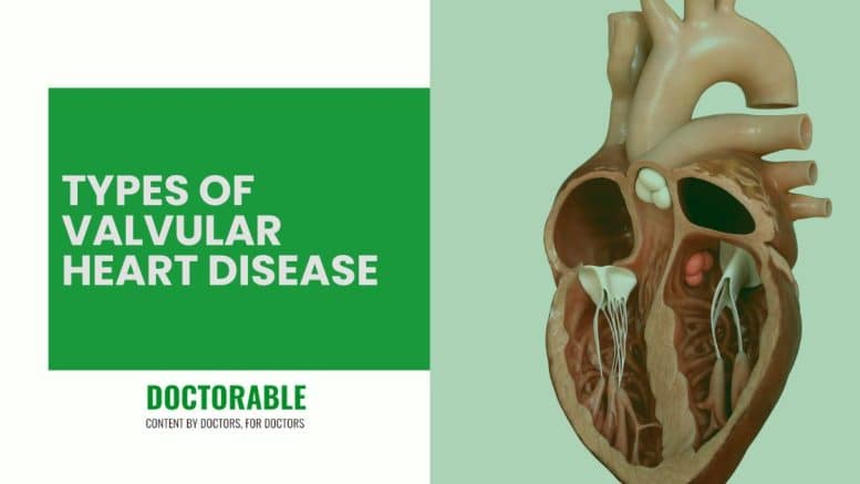
Types of Valvular Heart Disease
Valvular Stenosis
Heart valves can become rigid, leading to a narrowing of the valve opening and restricting blood flow, which is known as valve stenosis. This condition reduces the amount of blood that can flow through the valve and, in severe cases, it may eventually lead to systemic hemodynamic alterations.
- Mitral valve stenosis: stenosis of the mitral valve decreases blood flow from the left atrium to the left ventricle. The resulting pressure buildup in the left atrium can lead to its enlargement and even to pulmonary edema.
- Aortic valve stenosis: if the aortic valve gets narrowed, the blood that flows through the aorta from the heart to the rest of the body gets restricted, thus requiring the left ventricle to contract harder to compensate. In patients with aortic valve stenosis, the severity of the condition increases gradually over the course of the years, which leads to the development of left ventricular concentric hypertrophy. (5)
- Tricuspid valve stenosis: when stenosis of the tricuspid valve happens, the blood is unable to flow from the right atrium to the right ventricle, leading to subsequent enlargement of the atrium and increased pressure and blood flow in the contributing veins.
- Pulmonary valve stenosis: narrowing of the pulmonary valve impairs the blood flow from the right ventricle to the lungs through the pulmonary arteries.
Valvular Regurgitation
Regurgitation happens when the valve fails to close adequately, allowing the blood to leak backward and disrupting the normal one-directional blood flow in the heart.
- Mitral valve regurgitation: in mitral valve regurgitation the blood flows backward from the left ventricle into the left atrium. The excess exertion that the left ventricle needs to perform causes eccentric left-ventricular hypertrophy and leads to pulmonary edema in severe cases.
- Aortic valve regurgitation: aortic valve regurgitation occurs when the aortic valve fails to close properly during diastole, which causes the blood to flow backward from the aorta into the left ventricle. Over time, this can result in left ventricular dysfunction with eccentric hypertrophy, heart failure, and even sudden cardiac death. (6)
- Tricuspid valve regurgitation: this occurs when the tricuspid valve does not close properly, allowing blood to leak back from the right ventricle to the right atrium.
- Pulmonary valve regurgitation: when the pulmonary valve does not close adequately, blood from the pulmonary arteries flows back into the right ventricle, impeding the appropriate gas exchange and leading to Pulmonary Hypertension.
Valvular Prolapse
Valvular prolapse happens when the leaflets slip out of place in heart systole, which leads to improper closure of the valves and the subsequent disruption of normal blood flow, which can produce blood regurgitation.
- Mitral valve prolapse: mitral valve prolapse happens when the valve leaflets bulge into the atrium during systole, thus allowing blood to flow backward (regurgitate). Mitral valve prolapse is also known as Barlow’s syndrome, click-murmur syndrome, floppy valve syndrome, or billowing mitral valve syndrome. (6)
- Aortic, tricuspid, and pulmonary valve prolapse: prolapse in these valves happen less frequently than in the mitral valve, but the mechanism is the same, and normal one-direction blood flow in the heart gets disrupted.
The most frequent types of VHD are aortic stenosis, mitral regurgitation, mitral stenosis, and aortic regurgitation. (7)
Etiology and Pathophysiology of Valvular Heart Disease
The etiology and pathophysiology of valvular heart disease are multifactorial and complex, involving a combination of genetic, environmental, and lifestyle factors.
When VHD is present, the cardiac chambers experience either increased pressure or volume overload. This initiates a sequence of adaptive responses aimed at reducing wall tension and maintaining cardiac output, which include chamber hypertrophy and/or dilation. However, these compensatory mechanisms are followed by maladaptive changes in the myocardium, such as microvascular ischemia, reactive fibrosis, and cell death with fibrotic replacement. These alterations significantly impair left ventricular function, which gradually leads to a reduction in left ventricular ejection fraction (LVEF) produced over time. (6, 8)
VHD constitutes a major cause of morbidity and mortality, affecting around 2% of the world’s population, and it can be classified into two main categories: congenital or acquired. (9-11)
Congenital
Congenital VHD results from abnormalities in valve development during embryogenesis. Examples of congenital VHD include aortic stenosis, bicuspid aortic valve disease, pulmonary valve stenosis, and Marfan syndrome. (2, 4, 12)
Acquired
Acquired VHD results from a wide number of factors that can affect the valves after birth. The most common causes of acquired VHD are age-related degenerative changes, rheumatic fever, infective endocarditis, radiation therapy, drug use, and systemic diseases such as systemic lupus erythematosus. (2, 4)
Due to aging population, degenerative valve disease is the most common form of VHD in the United States and other developed countries. Degenerative VHD is characterized by progressive fibrosis, calcification, and thickening of the valve leaflets, resulting in valvular stenosis or regurgitation. (10, 13-15)
In developing countries, rheumatic heart disease accounts for most heart valve pathologies. Rheumatic fever, which is caused by group A streptococcal infections, can lead to VHD through the formation of immune complexes that produce inflammation and scarring of the valve leaflets. (10, 13, 16, 17)
Clinical Findings in Valvular Heart Disease
Depending on the severity of VHD, patients may present with a range of symptoms such as dyspnea, fatigue, chest pain, syncope, and/or palpitations. Nevertheless, symptoms of VHD frequently appear at a late stage, when myocardial damage has already occurred, and systemic and pulmonary circulation has been compromised. Therefore, in most cases, patients may be suspected to have VHD based on incidental findings of valvular abnormalities shown in noninvasive testing. (14, 18)
Since decisions about treatment are based on the presence or absence of symptoms, a throughout medical history and physical examination is crucial in the evaluation of patients with VHD.
However, assessment of symptoms can be challenging due to the slow and progressive nature of most valve lesions which leads patients not to recognize or report common symptoms because they have already normalized them as part of their daily life. Furthermore, symptoms may also be related to concomitant comorbidities, such as obesity, pulmonary disease, or coronary artery disease, especially in elderly patients. (7, 19)
In any case, severe VHD should always be suspected in patients who have a history of prior embolism or pulmonary edema, and in patients with pulmonary hypertension, cardiomegaly, or atrial fibrillation, regardless of their symptoms. (7, 19)
The following table summarizes the most common physical exam findings suggestive of valve disease: (2 ,5 ,7, 14, 16, 20)
| Type of VHD | Common Findings |
|---|---|
| Aortic Stenosis | Crescendo-decrescendo systolic murmur. Normal or soft S1. Single or paradoxically split S2. Palpable and audible S4. Pulsus parvus et tardus (may not be present in elderly patients). Prominent a waves may be present in the jugular venous pulse. |
| Aortic Regurgitation | High-frequency decrescendo diastolic murmur. Water-hammer or Corrigan pulse. Bisferiens pulse may be palpable. Increased systolic blood pressure with low diastolic blood pressure. “Pistol shot” sounds over the femoral arteries. Capillary pulsations visible at the fingertips, lips, and tongue. |
| Mitral Stenosis | Accentuated S1. Opening snap followed by a diastolic rumble, heard best in left lateral decubitus position. |
| Mitral Regurgitation | Loud early systolic murmur or holosystolic murmur. Widely split S2. S3 and diastolic rumble may be present. |
| Mitral Valve Prolapse | Late systolic murmur and midsystolic clicks. |
Diagnosis of Valvular Heart Disease
The main test used for diagnosing and evaluating valvular heart disease is a comprehensive transthoracic echocardiogram with doppler.
Other tests such as electrocardiography, chest radiography, cardiac computed tomography, cardiac magnetic resonance, stress testing, and diagnostic hemodynamic cardiac catheterization may be further required to determine adequate treatment and evaluate the possible surgical risk for a patient with VHD.
Echocardiography
Two-dimensional transthoracic echocardiography (TTE) with doppler is considered the test of choice in the initial and repeat evaluation of patients with suspected or confirmed VHD as it provides valuable information about the morphology and function of the cardiac chambers, valves, and great vessels. (6, 7, 14, 21, 22)
TTE (and, in some instances, transesophageal echocardiography, or TEE) is useful to help diagnose, assess the severity, determine the etiology, evaluate hemodynamic changes, and detect other associated valve lesions in patients with VHD. (6, 14, 16, 23, 24)
Electrocardiography
Electrocardiography (ECG) findings may be normal in the early stages or may show signs of chamber hypertrophy or dilation.
Depending on the specific type of VHD, it might be common to find repolarization or conduction abnormalities, including left and right bundle branch block, atrial fibrillation, or a leftward or rightward axis deviation, among other anomalies. (16)
Chest Radiography
A chest radiography can be useful in the initial examination of patients with known or suspected VHD to assess cardiac size and aorta changes, as well as evaluate the presence of pulmonary vascular congestion and other associated lung pathologies. (16)
Cardiac computed tomography
Cardiac computed tomography (CT) may be a useful tool to assess valve calcification as well as evaluate the presence and severity of aortic root and ascending aortic dilatation in patients with concomitant aortic aneurysms. (16, 19)
Cardiac magnetic resonance imaging
Cardiac magnetic resonance imaging (CMR) can be used to measure the anatomic valve area and assess the degree of regurgitation, if present. This test can also be useful to evaluate the morphology of the aorta and cardiac chambers. However, in routine practice, the use of CMR is limited due to its availability and elevated cost. (7, 16, 19, 25, 26)
Stress Testing
A stress test is useful to determine functional capacity, especially in patients who have a discrepancy in symptoms and resting measures of left ventricular function and pulmonary artery pressure.
Since VHD has a very slow progression rate, many patients may deny symptoms because they are unable to recognize them as such, given that they gradually limited their lifestyle over the years. Therefore, these patients may benefit from a stress test since it will help identify those who are truly symptomatic.
However, this test should not be performed on patients with unequivocal symptoms.
Performing a doppler TTE before and after a stress test can provide additional information about the hemodynamic status of the patient, especially when surgical intervention is being considered as a possible treatment. (5, 7, 16, 19)
Diagnostic hemodynamic cardiac catheterization
Hemodynamic cardiac catheterization is primarily used before a surgical intervention to identify and assess the presence of associated coronary artery disease (CAD) in certain patients with the appropriate age and risk factor profile.
However, diagnostic cardiac catheterization can also be indicated in limited cases to define hemodynamic abnormalities when the severity of valve stenosis or regurgitation cannot be ascertained due to discrepancies between clinical and non-invasive imaging test findings, especially in symptomatic patients. (2, 7, 19)
Treatment and Prevention
Treatment of VHD depends on the severity of the condition, the valve(s) affected, and the patient’s age and overall health status.
Medical therapy may include diuretics, beta-adrenergic blockers, angiotensin-receptor blockers (ARBs), or angiotensin-converting enzyme (ACE) inhibitors in those patients with other risk factors for CAD and for which medical therapy may help delay the progression of VHD. (14, 15, 19, 27)
Prophylaxis for infective endocarditis should be given to all patients with significant valve disease, particularly in cases with regurgitant valve lesions. While, prophylaxis for rheumatic fever is indicated in young patients with evidence of rheumatic etiology, especially in countries with a high prevalence of rheumatic disease. (5, 14, 19, 28)
Surgical intervention, such as valve replacement with either mechanical or biological prostheses, or valve repair, is considered to be the treatment of choice in patients who develop symptoms and in those who are asymptomatic with left ventricular dysfunction. However, in truly asymptomatic patients, the indications for surgery should be individualized taking into consideration the balance of risks and benefits and the patient’s wishes. (7, 14, 29-34)
Percutaneous balloon valvotomy may be a reasonable option for highly symptomatic patients who are not surgical candidates and for young patients with noncalcified valves; however, this procedure has many limitations. (16, 35)
Preventive measures involve the management of any underlying conditions that may increase the risk of developing VHD, including hypertension, hypercholesterolemia, and type II diabetes mellitus; as well as keeping healthy lifestyle habits, such as maintaining an adequate weight, exercising regularly, and cessation of tobacco, alcohol, and caffeine. (2, 3, 14)
Disclosures
The author does not report any conflict of interest.
Disclaimer
This information is for educational purposes and is not intended to treat disease or supplant professional medical judgment. Physicians should follow local policy regarding the diagnosis and management of medical conditions.
See Also
Heart Failure with Preserved Ejection Fraction
Dyspnea Due to Respiratory Causes
References
- How the Heart Works – How Blood Flows through the Heart. NHLBI, NIH [Internet]. www.nhlbi.nih.gov. 2022. Available from: https://www.nhlbi.nih.gov/health/heart/blood-flow
- Bonow RO, Mann DL, Zipes DP, Libby P. Braunwald’s Heart Disease E-Book: A Textbook of Cardiovascular Medicine [Internet]. Google Books. Elsevier Health Sciences; 2011 [cited 2023 Mar 19]. Available from: https://books.google.com.ar/books?hl=en&lr=&id=b5wADkB9oDoC&oi=fnd&pg=PP1&ots=VmZl2ejyFt&sig=saweIlue4g5rTuK0BnTAcaT39Mk&redir_esc=y#v=onepage&q&f=false
- Zeng Y, Sun R, Li X, Liu M, Chen S, Zhang P. Pathophysiology of valvular heart disease. Experimental and Therapeutic Medicine [Internet]. 2016 Feb 5 [cited 2023 Mar 19];11(4):1184–8. Available from: https://www.ncbi.nlm.nih.gov/pmc/articles/PMC4812598/
- Boudoulas KD, Borer JS, Boudoulas H. Etiology of Valvular Heart Disease in the 21st Century. Cardiology. 2013;126(3):139–52. Available from: https://www.karger.com/Article/FullText/354221
- Bonow RO, Carabello BA, Chatterjee K, de Leon AC, Faxon DP, Freed MD, et al. 2008 Focused update incorporated into the ACC/AHA 2006 guidelines for the management of patients with valvular heart disease: a report of the American College of Cardiology/American Heart Association Task Force on Practice Guidelines (Writing Committee to Revise the 1998 Guidelines for the Management of Patients With Valvular Heart Disease): endorsed by the Society of Cardiovascular Anesthesiologists, Society for Cardiovascular Angiography and Interventions, and Society of Thoracic Surgeons. Circulation [Internet]. 2008 [cited 2023 Mar 19];118(15):e523-661. Available from: https://www.ncbi.nlm.nih.gov/pubmed/18820172
- Ajmone Marsan N, Delgado V, Shah DJ, Pellikka P, Bax JJ, Treibel T, et al. Valvular heart disease: shifting the focus to the myocardium. European Heart Journal [Internet]. 2022 Sep 28 [cited 2023 Mar 20];44(1):28–40. Available from: https://academic.oup.com/eurheartj/article/44/1/28/6724464#392708002
- Lung B. Recommendations on the management of the asymptomatic patient with valvular heart disease. European Heart Journal. 2002 Aug 15;23(16):1253–66.Available from: https://www.researchgate.net/profile/Christophe-Tribouilloy/publication/10801539_Recommendations_on_the_management_of_the_asymptomatic_patient_with_valvular_heart_disease/links/0912f50c792e81650b000000/Recommendations-on-the-management-of-the-asymptomatic-patient-with-valvular-heart-disease.pdf
- Carabello BA. How Does the Heart Respond to Aortic Stenosis. Circulation: Cardiovascular Imaging. 2013 Nov;6(6):858–60. Available from: https://www.ahajournals.org/doi/full/10.1161/circimaging.113.001242
- Nkomo VT, Gardin JM, Skelton TN, Gottdiener JS, Scott CG, Enriquez-Sarano M. Burden of valvular heart diseases: a population-based study. Lancet (London, England) [Internet]. 2006;368(9540):1005–11. Available from: https://www.ncbi.nlm.nih.gov/pubmed/16980116
- Coffey S, Roberts-Thomson R, Brown A, Carapetis J, Chen M, Enriquez-Sarano M, et al. Global epidemiology of valvular heart disease. Nature Reviews Cardiology [Internet]. 2021 Jun 25;1–12. Available from: https://www.nature.com/articles/s41569-021-00570-z
- Go AS, Mozaffarian D, Roger VL, Benjamin EJ, Berry JD, Borden WB, et al. Heart Disease and Stroke Statistics-2013 Update: A Report From the American Heart Association. Circulation [Internet]. 2012;127(1):e6–245.
- Le Gloan L, Mercier LA, Dore A, Marcotte F, Ibrahim R, Mongeon FP, et al. Recent Advances in Adult Congenital Heart Disease. Circulation Journal. 2011;75(10):2287–95. Available from: https://pubmed.ncbi.nlm.nih.gov/21881245/
- Lung B, Delgado V, Rosenhek R, Price S, Prendergast B, Wendler O, et al. Contemporary Presentation and Management of Valvular Heart Disease. Circulation. 2019 Oct;140(14):1156–69. Available from: https://pubmed.ncbi.nlm.nih.gov/31510787/
- Nishimura RA, Otto CM, Bonow RO, Carabello BA, Erwin JP, Guyton RA, et al. 2014 AHA/ACC Guideline for the Management of Patients With Valvular Heart Disease. Circulation. 2014 Jun 10;129(23). Available from: https://www.ahajournals.org/doi/full/10.1161/CIR.0000000000000031#d1e1603
- Boudoulas H, Wooley CF. Floppy mitral valve, mitral valve prolapse, and mitral valvular regurgitation. Current Treatment Options in Cardiovascular Medicine. 2001 Feb;3(1):15–24. Available from: https://pubmed.ncbi.nlm.nih.gov/11139786/
- Maganti K, Rigolin VH, Sarano ME, Bonow RO. Valvular Heart Disease: Diagnosis and Management. Mayo Clinic Proceedings [Internet]. 2010 May;85(5):483–500. Available from: https://www.ncbi.nlm.nih.gov/pmc/articles/PMC2861980/
- Carapetis JR, Steer AC, Mulholland EK, Weber M. The global burden of group A streptococcal diseases. The Lancet Infectious diseases [Internet]. 2005;5(11):685–94. Available from: https://www.ncbi.nlm.nih.gov/pubmed/16253886
- Ross J, Braunwald E. Aortic Stenosis. Circulation. 1968 Jul;38(1s5). Available from: https://pubmed.ncbi.nlm.nih.gov/4894151/
- Vahanian A, Alfieri O, Andreotti F, Antunes MJ, Barón-Esquivias G, Baumgartner H, et al. Guidelines on the management of valvular heart disease (version 2012). European Heart Journal [Internet]. 2012 Aug 24 [cited 2023 Mar 21];33(19):2451–96. Available from: https://academic.oup.com/eurheartj/article/33/19/2451/483360
- Shipton B, Wahba H. Valvular Heart Disease: Review and Update. American Family Physician [Internet]. 2001 Jun 1;63(11):2201–9. Available from: https://www.aafp.org/pubs/afp/issues/2001/0601/p2201.html
- Lancellotti P, Tribouilloy C, Hagendorff A, Moura L, Popescu BA, Agricola E, et al. European Association of Echocardiography recommendations for the assessment of valvular regurgitation. Part 1: aortic and pulmonary regurgitation (native valve disease). European Journal of Echocardiography [Internet]. 2010 Apr 1 [cited 2023 Mar 21];11(3):223–44. Available from: https://www.escardio.org/static_file/Escardio/education/eLearning/webinars/general-cardiology/eae-recommendations-assessment-valvular-regurgitation.pdf
- Zamorano JL, Bax JJ, Rademakers FE, Knuuti J, editors. The ESC Textbook of Cardiovascular Imaging [Internet]. Google Books. Oxford University Press; 2014 [cited 2023 Mar 21]. Available from: https://books.google.com.ar/books?hl=en&lr=&id=_fAkBwAAQBAJ&oi=fnd&pg=PP1
- Enriquez-Sarano M, Avierinos JF, Messika-Zeitoun D, Detaint D, Capps M, Nkomo V, et al. Quantitative Determinants of the Outcome of Asymptomatic Mitral Regurgitation. New England Journal of Medicine. 2005 Mar 3;352(9):875–83. Available from: https://pubmed.ncbi.nlm.nih.gov/15745978/
- Zoghbi W. Recommendations for evaluation of the severity of native valvular regurgitation with two-dimensional and doppler echocardiography. Journal of the American Society of Echocardiography. 2003 Jul;16(7):777–802. Available from: https://pubmed.ncbi.nlm.nih.gov/12835667/
- Kilner PJ, Manzara CC, Mohiaddin RH, Pennell DJ, Sutton MG, Firmin DN, et al. Magnetic resonance jet velocity mapping in mitral and aortic valve stenosis. Circulation. 1993 Apr;87(4):1239–48. Available from: https://pubmed.ncbi.nlm.nih.gov/8462150/
- Roes SD, Hammer S, van der Geest RJ, Marsan NA, Bax JJ, Lamb HJ, et al. Flow assessment through four heart valves simultaneously using 3-dimensional 3-directional velocity-encoded magnetic resonance imaging with retrospective valve tracking in healthy volunteers and patients with valvular regurgitation. Investigative Radiology [Internet]. 2009 Oct 1 [cited 2023 Mar 22];44(10):669–75. Available from: https://pubmed.ncbi.nlm.nih.gov/19724233/
- Lin M, Chiang HT, Lin SL, Chang MS, Chiang BN, Kuo HW, et al. Vasodilator therapy in chronic asymptomatic aortic regurgitation: Enalapril versus hydralazine therapy. Journal of the American College of Cardiology. 1994 Oct;24(4):1046–53. Available from: https://www.ncbi.nlm.nih.gov/pmc/articles/PMC2861980/
- Wilson W, Taubert KA, Gewitz M, Lockhart PB, Baddour LM, Levison M, et al. Prevention of Infective Endocarditis. Circulation. 2007 Oct 9;116(15):1736–54. Available from: https://www.ahajournals.org/doi/full/10.1161/CIRCULATIONAHA.106.183095#d1e1015
- Bouma B, van den Brink RBA, van der Meulen JHP, Verheul H, Cheriex E, Hamer H, et al. To operate or not on elderly patients with aortic stenosis: the decision and its consequences. Heart [Internet]. 1999 Aug 1 [cited 2023 Mar 22];82(2):143–8. Available from: https://www.ncbi.nlm.nih.gov/pmc/articles/PMC1729124/
- Bach DS, Siao D, Girard SE, Duvernoy C, McCallister BD, Gualano SK. Evaluation of patients with severe symptomatic aortic stenosis who do not undergo aortic valve replacement: the potential role of subjectively overestimated operative risk. Circulation Cardiovascular Quality and Outcomes [Internet]. 2009 Nov 1;2(6):533–9. Available from: https://pubmed.ncbi.nlm.nih.gov/20031890/
- Rosenhek R, Rader F, Klaar U, Gabriel H, Krejc M, Kalbeck D, et al. Outcome of Watchful Waiting in Asymptomatic Severe Mitral Regurgitation. Circulation. 2006 May 9;113(18):2238–44. Available from: https://pubmed.ncbi.nlm.nih.gov/16651470/
- Enriquez-Sarano M, Tajik AJ. Aortic Regurgitation. New England Journal of Medicine. 2004 Oct 7;351(15):1539–46. Available from: https://pubmed.ncbi.nlm.nih.gov/15470217/
- Klodas E, Enriquez-Sarano M, Tajik AJamil, Mullany CJ, Bailey KR, Seward JB. Aortic regurgitation complicated by extreme left ventricular dilation: Long-term outcome after surgical correction. Journal of the American College of Cardiology. 1996 Mar;27(3):670–7. Available from: https://pubmed.ncbi.nlm.nih.gov/8606280/
- Chaliki HP, Mohty D, Avierinos JF, Scott CG, Schaff HV, Tajik AJ, et al. Outcomes After Aortic Valve Replacement in Patients With Severe Aortic Regurgitation and Markedly Reduced Left Ventricular Function. Circulation. 2002 Nov 19;106(21):2687–93. Available from: https://pubmed.ncbi.nlm.nih.gov/12438294/
- Lieberman EB, Bashore TM, Hermiller JB, Wilson JS, Pieper KS, Keeler GP, et al. Balloon aortic valvuloplasty in adults: failure of procedure to improve long-term survival. Journal of the American College of Cardiology [Internet]. 1995 Nov 15;26(6):1522–8. Available from: https://pubmed.ncbi.nlm.nih.gov/7594080/